3D Oct scanning for the cornea and anterior eye is currently in its infancy. The OCT scanning machines are relatively new technology and had initially been developed to investigate the retina, at the back of the eye. It is only within the last few years that the potential for investigation of the anterior eye and cornea has been realised.
It works by reflecting infra-red light off various structures within the eye and measuring how much light is reflected. By detailed analysis of this reflected pattern of light the software can to rebuild a three dimensional model of the internal ocular structure such as the cornea. This gives a highly detailed perspective of the eye and an understanding of what is actually happening to the corneal shape and curvature of the cornea in keratoconus. The information is of great value to a contact lens practitioner as it can also be used post lens fitting to assess how a lens is performing on the eye. It also eludes to the severity the keratoconus/ectasia and how best to treat it.
On this page I have given some examples of cornea shapes to help illustrate what affect the different conditions can have on the cornea and to explain a little why complex lenses are required.
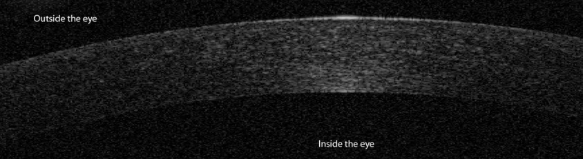
The curvature of a normal eye
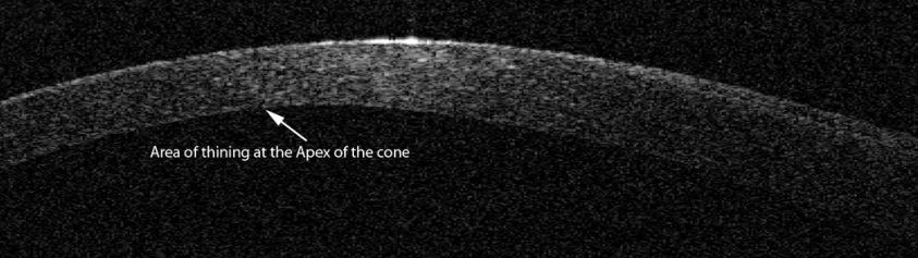
A keratoconic eye, the cornea buldges into a cone due to the thinning of the corneal tissue seen here
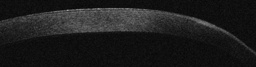
PMD, essentially the same process as keratoconus although the corneal thinning is much more peripheral as see here
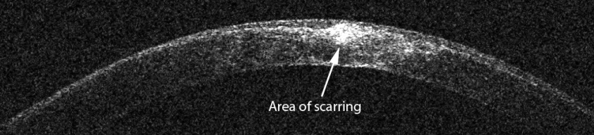
In keratoconus the cornea can scar at the apex of the cone as seen here
